Biological Products:
Bioaugmentation products for Wastewater applications in Papermills, Refineries, Chemical, Tanneries, Municipalities, Textiles, Steel, Agriculture, Animal feedlot, Gun Powder plant, Food and Beverage- Dairy Products, Orange Juice factory, Wineries, Cookie factory, Vegetable processing plant, Meat packing, Barbecue Restaurant, Aquaculture, Ornamental Ponds with algae , CAFO, Nursing homes, Military, Campgrounds, Universities, Regulatory agencies, River and Lake remediation
Lab Services:
Filamentous Identification Lab Service. One reason to identify filaments is to determine the filaments characteristics and then determine the type present. If the type is found out, a root cause can usually be associated with a particular filament. If the cause is known, then a correction can be made to alleviate problems. Chlorination is only a quick fix. Without process changes, filaments will grow back after chlorination. Wastewater Biomass Analyses and Cooling Tower Analyses also available
Training Materials:
Training is an integral part of any job. Not everyone is at the same level of training. Many people want beginning concepts and basics. Some need technical information or troubleshooting. Some want equipment, technology or process information. We have developed a full set of Basic training, Advanced training, Filamentous Identification the Easy Way as well as custom training CD's Manuals. We also provide hands-on training classes and soon will have an Online "E-University".
|
Microscope Bacterial Slides - Pre-Made Slide Sets From Various Wastewater Treatment
Latest News!
What's New!
We have just added "Virtual Audits" to our capabilities. Check out our new Services. We are in the process of developing new courses for our ""Online E-University" in order to meet the needs of our global customers that cannot travel to our public classes.Visit our new website www.WastewaterElearning.com/Elearning
Check with us for the next Filamentous ID 2 Day Training Class
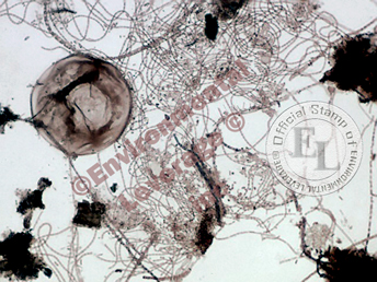
We have been asked by numerous customers to sell them some of the slides we used in our hands on Filamentous Identification training classes. So we created a new item for you to practice with your own microscope. It is one thing to look at photos of Gram and Neisser stains, it is another to try to find the filaments on a stain. They do not always pose and sit still for you. Also, we tend to "crop" the photos we take so that you can focus on just the filaments we are demonstrating.
Our Lab at Environmental Leverage has created a set of Microscope Gram and Neisser slides. There are 10 sets from 10 different plants. Each slide can be used again and again for practice. The slides are labeled with the major filaments present. This is an outstanding set of slides. Great Identification tool for your lab.
In this case you will find a matching set of slides for each sample that we tested. There are samples from wastewater plants in many environments. You may find a sample from many Municipalities, some from food plants or Industrial settings and occasionally a pond or lagoon.
Each slide will have a thick spot and a smeared spot on the slide. We do that during slide preparation so that in case the sample has changes in the floc, high Zooglea present, slime due to septicity or in case of over or under colorization, there will be a “back-up” area stained.
Usually, we focus on the thinly smeared portion of the slide. Gram stains will have a G in the bottom corner of the slide and Neisser stains will have an N. The slides are numbered so you can put them back in the correct slot. There is also an internal ID number on the case, which lets us know which batch the slides are from. Gram stains have two colors- Gram negative is pink and Gram positive is purple. Keep in mind septicity in a plant can make filaments pick up excess coloring though. Place slide right side up; place one drop of immersion oil directly on the slide. Use 1000x bright field to examine the slide. Try to find the filaments at the outer edge of the floc structure or in a clearer spot. Remember, even though it appears that this is 2 dimensional, it still technically is 3 dimensional. In areas where the floc and filaments combine too thick, it may be harder to see the exact filament present.
Microscope Slide Set. 20 slides
with 10 sets.
|


